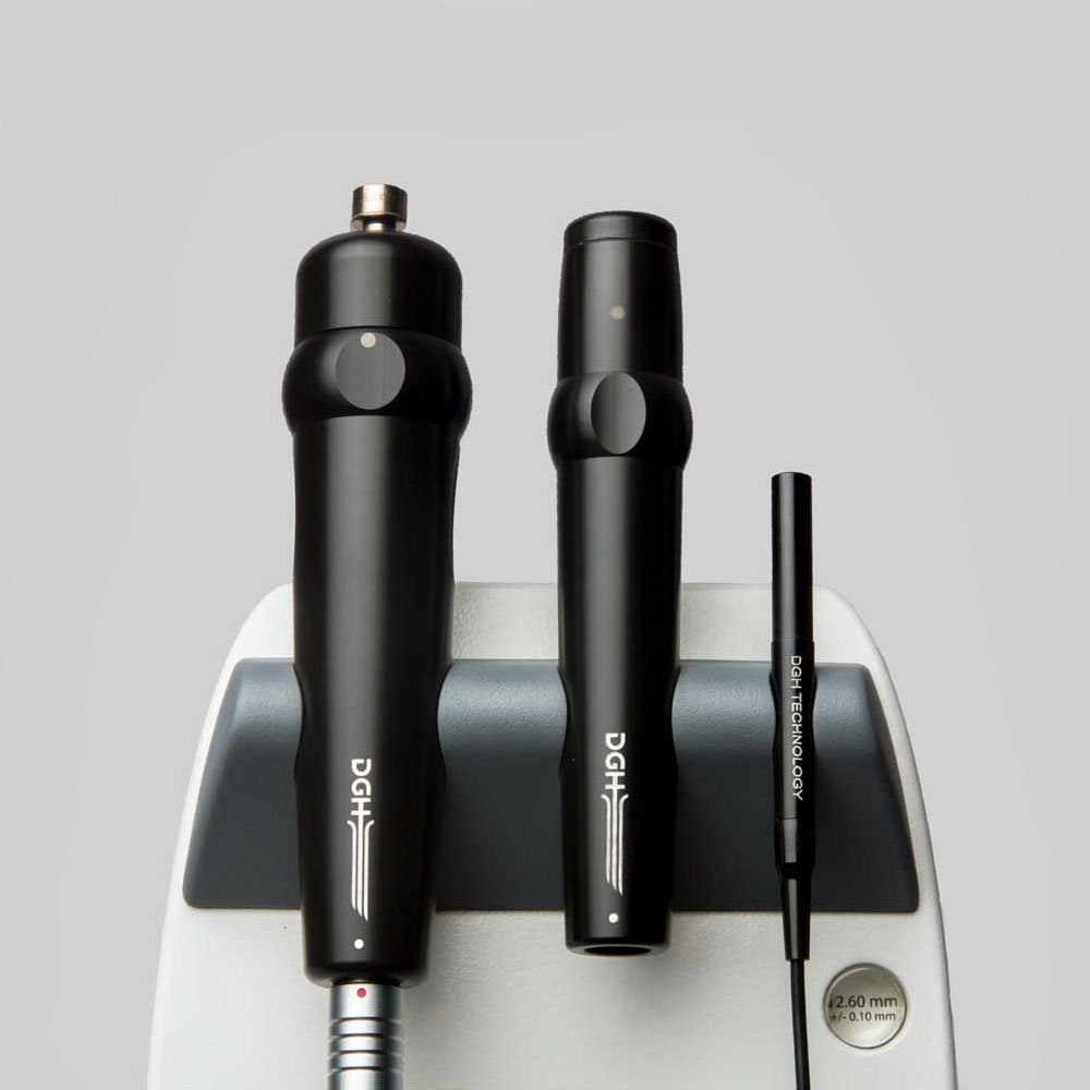
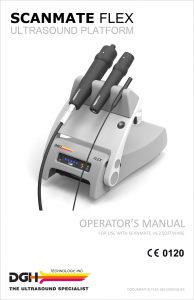
SCANMATE FLEX
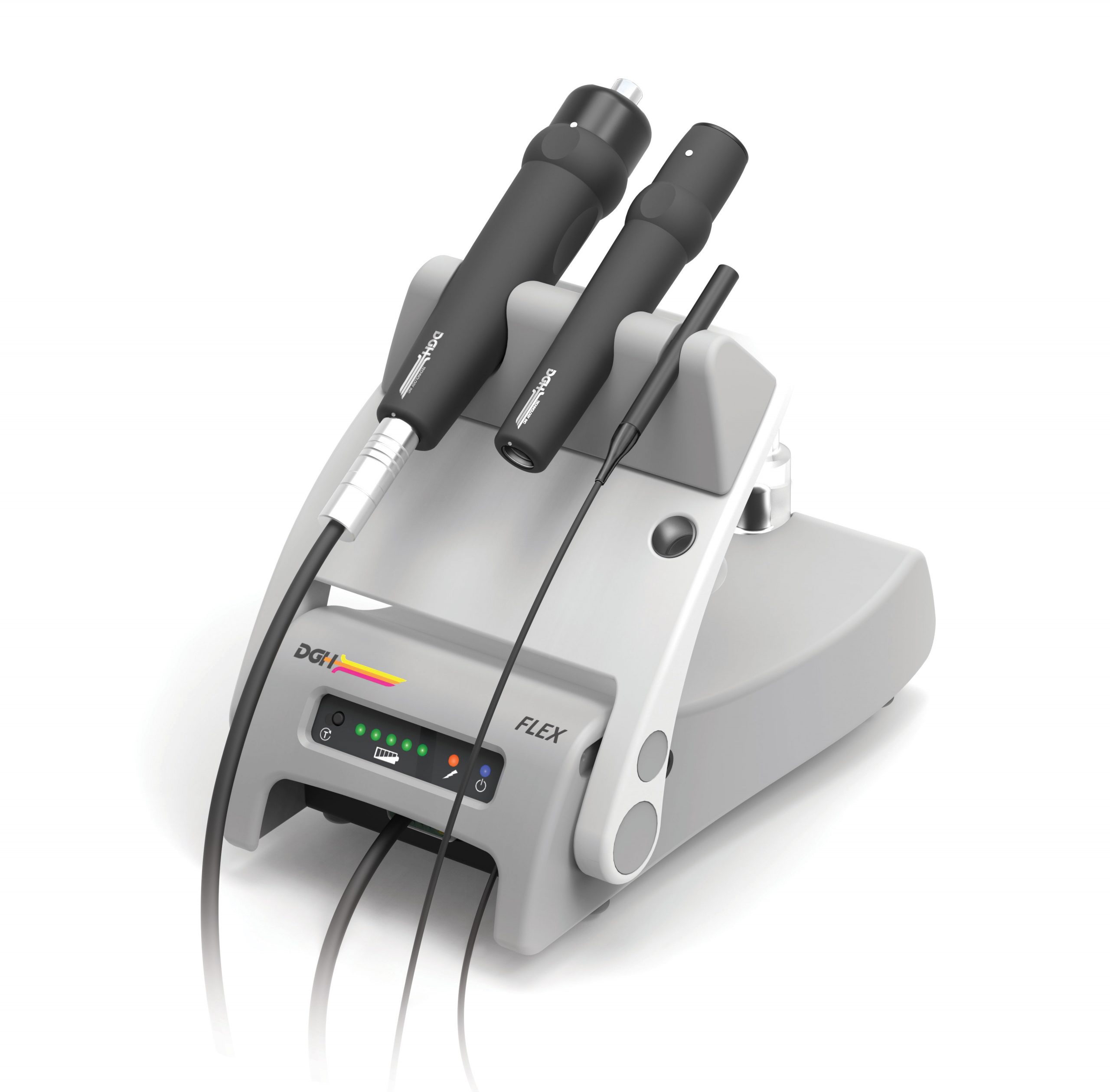
THREE PROBE OPTIONS, ONE POWERFUL PLATFORM
Maximize your technology investment by configuring the Scanmate Flex to meet your needs
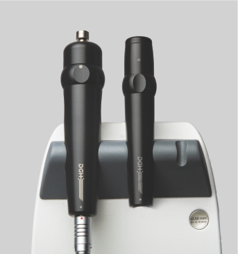
The Scanmate Flex, a portable, ophthalmic ultrasound platform, is the perfect product to enhance your practice’s diagnostic capabilities. The Flex provides any desired combination of A-Scan, B-Scan, and UBM. Its hallmark is flexibility.
A-Scan
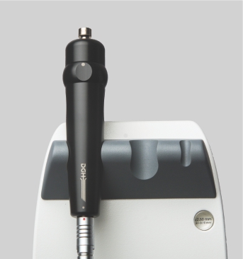
The Flex A-Scan uses DGH’s proven alignment algorithm to obtain repeatable and accurate axial length measurements. The IOL calculator is easy to use and includes most modern and post refractive formulas.
B-Scan
The redesigned B-Scan Probe, new with the Flex, provides clear imaging of the posterior segment of the eye, even when optical clarity is compromised.
UBM
The Flex UBM probe is indispensable when obtaining high resolution images of the anterior segment of the eye, including images of structures concealed by the iris or corneal opacities.
TOTAL IMAGING SOLUTION
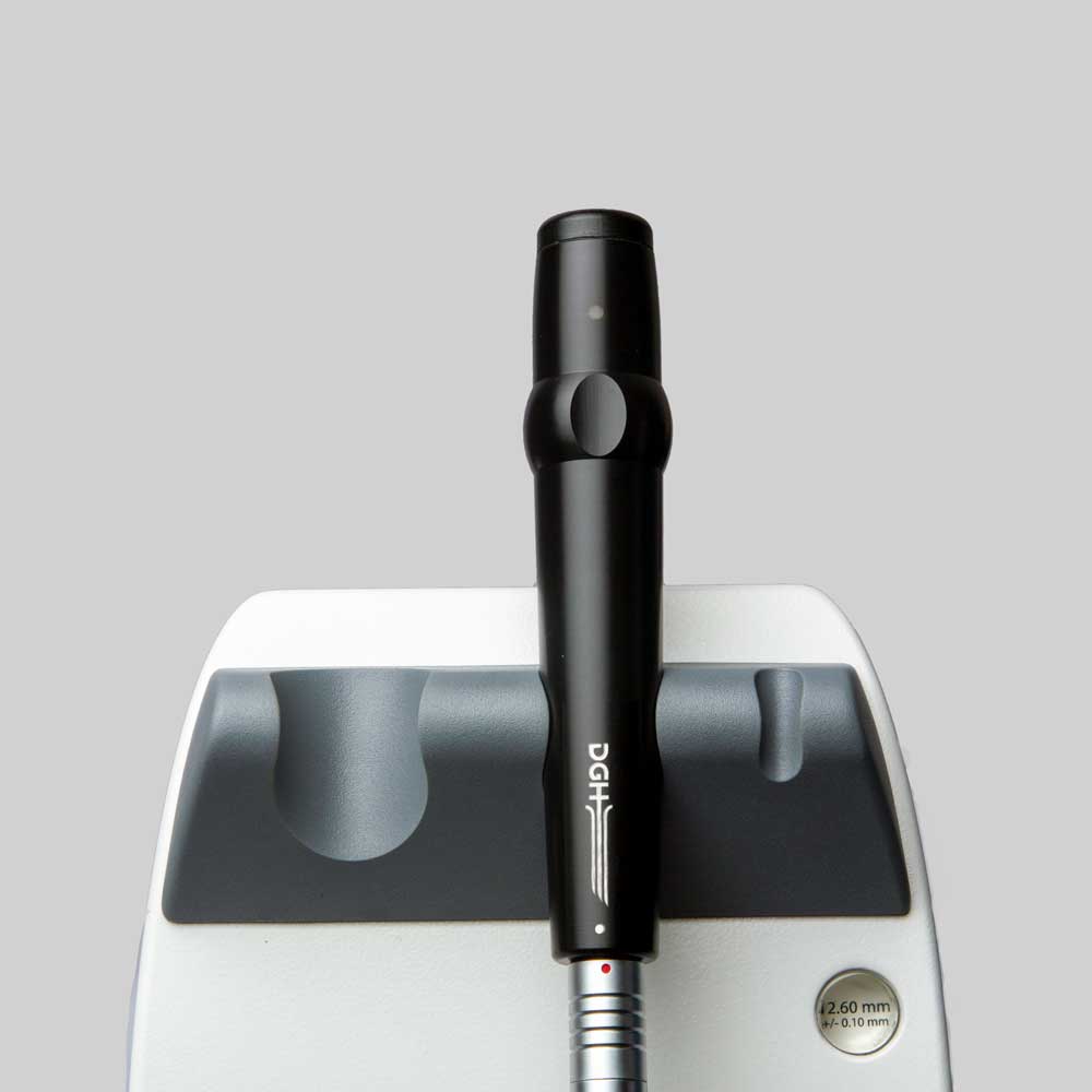
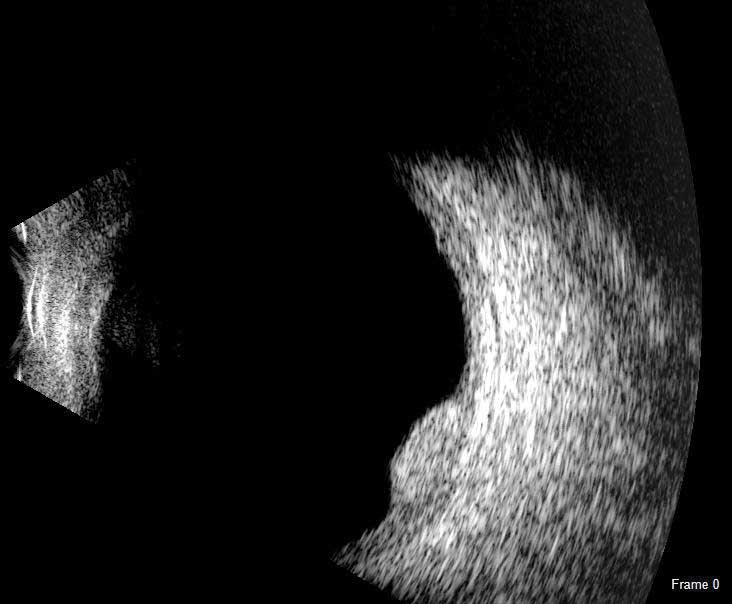
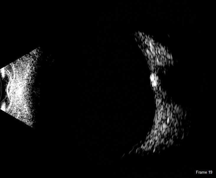
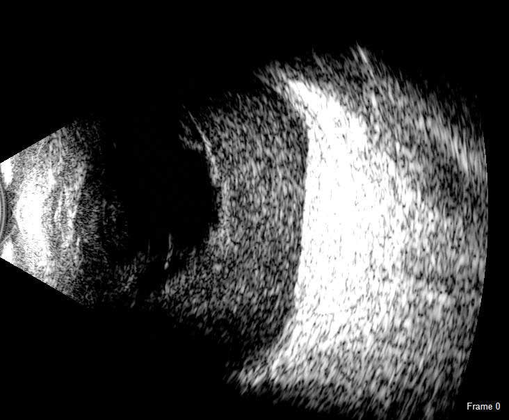
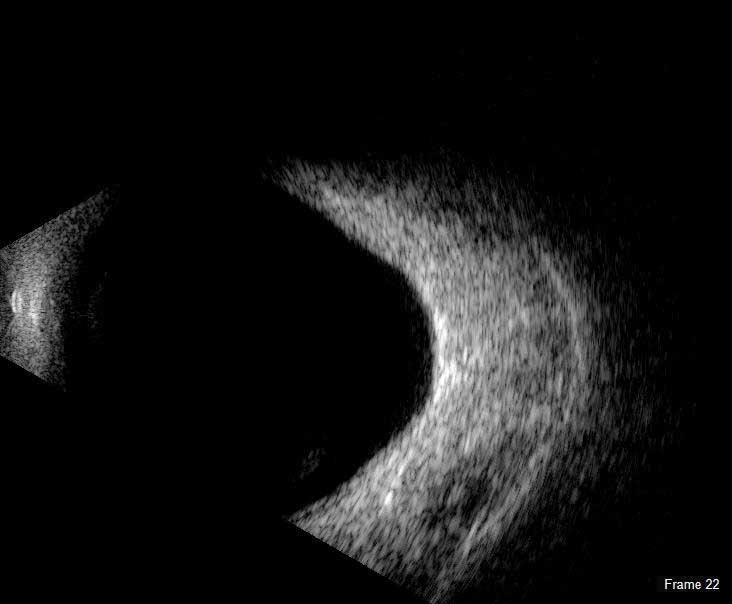
The Scanmate Flex B-Scan probe enables clinicians to capture clear and precise images and videos of the posterior segment of the eye. Ultrasonic B-scans are effective, even when opacities (such as dense cataract, blood, or anatomical structures) are present which obscure optical technologies.
The B-Scan probe is available in both 12.5 MHz and 20 MHz frequencies. Among the on-screen tools are calipers to measure structures, an area measurement tool and an annotation tool that gives you a way to indicate pathologies on the image.
B-Scan Diagnostic Applications
The Flex B-Scan delivers clear images of the posterior portion, even when optical clarity is compromised. B-Scan imaging can aid the evaluation of:
- Retinal Detachments
- Vitreous Detachments
- Vitreous Humor Pathologies
- Staphylomas
- Posterior Segment Pathologies
- Choroidal Pathologies
- Optic Nerve Pathologies
- Scleral Thickening
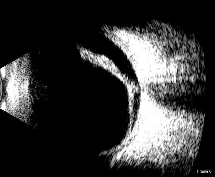
TOTAL IMAGING SOLUTION
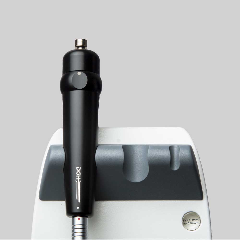
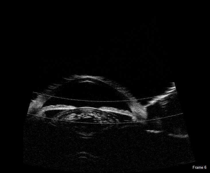
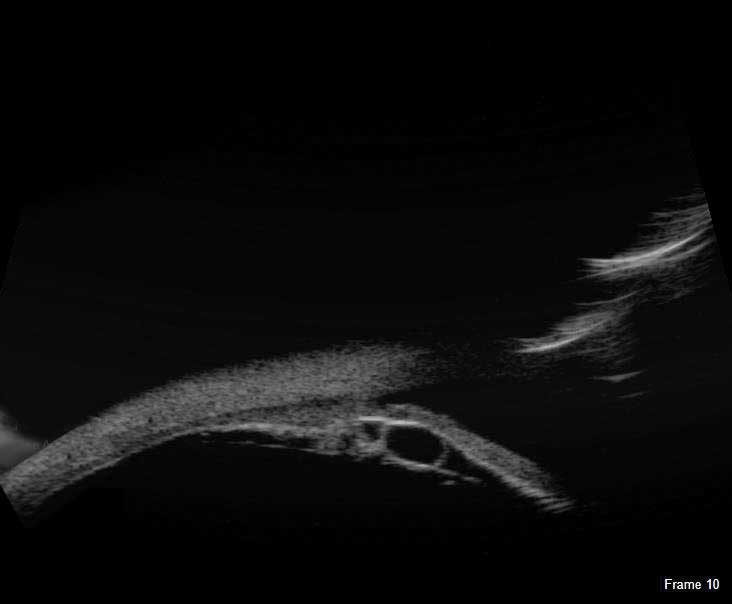
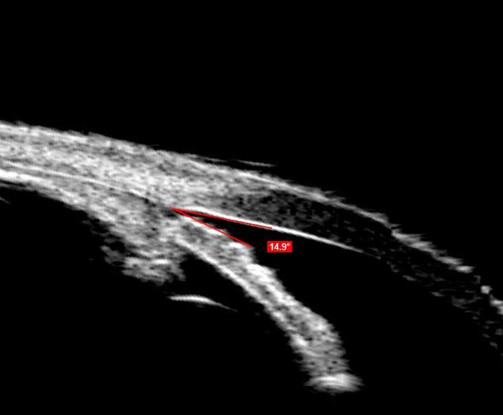
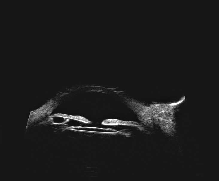
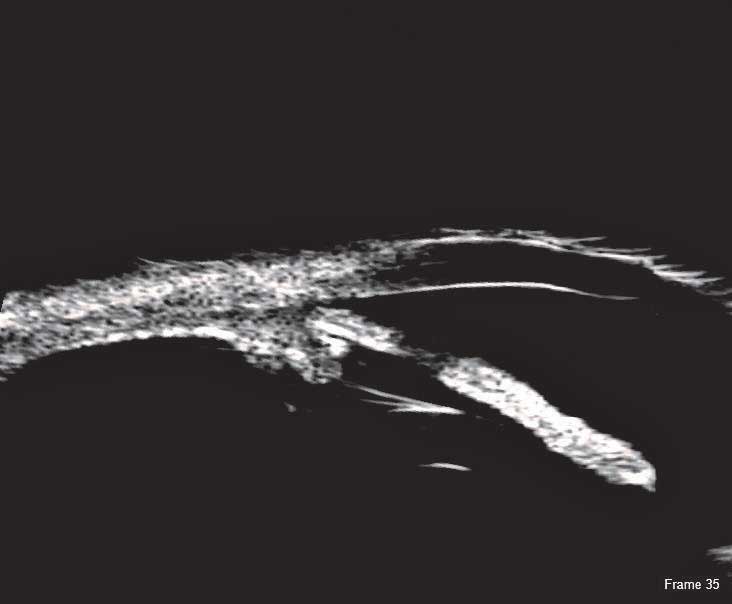
The UBM is indispensable when imaging the anterior segment. High-resolution UBM images provide the ability to observe structures concealed by the iris or corneal opacities.
Typical applications of the high resolution images obtained with either a 35 MHz or 50 MHz transducer include sulcus-to-sulcus measurement, angle-closure, and anterior chamber pathologies. The Scanmate software provides tools for angle, area and length measurement along with an annotation tool for indicating pathologies. A water-filled single use sterile Clear Scan® Probe cover is the only component that touches the eye.
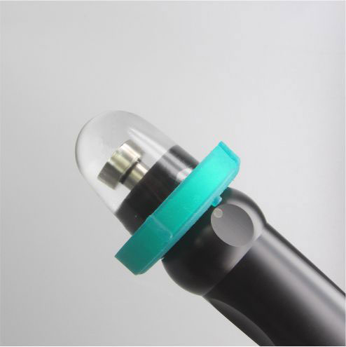
UBM Diagnostic Applications
The Flex UBM delivers clear images for the anterior segment, and the technology is presently being utilized in procedures such as:
- ACA Measurements
- Post-Lasik Corneal Evaluation
- IOL Position Monitoring
- Pre-Operative ICL Evaluation
- Diagnosis of Iris and Ciliary Cysts
- Glaucoma Post Surgery Evaluations
TOTAL IMAGING SOLUTION
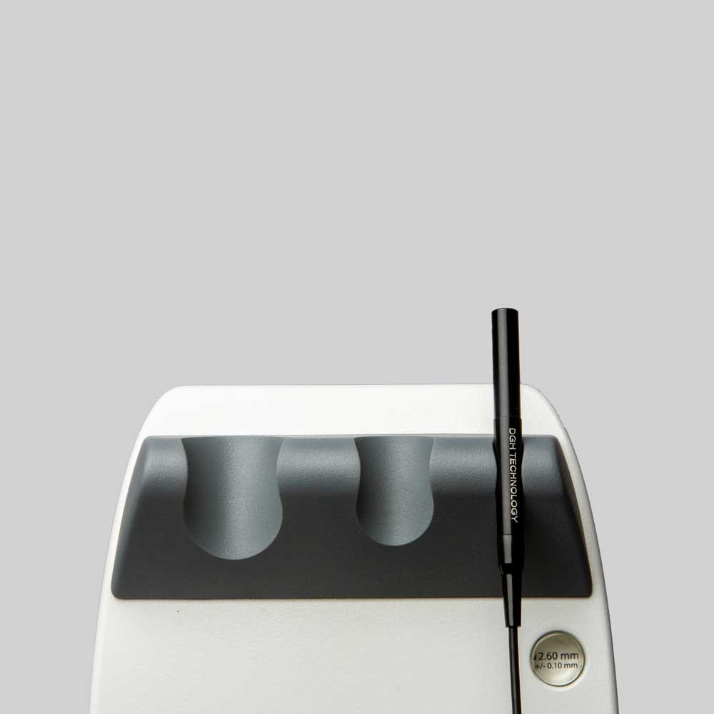
The innovative Flex A-Scan offers clinicians an unmatched level of usability and accuracy. Unique measurement guidance features assist in achieving optimal measurement values. These features allow the user to focus on application technique while the device’s software performs real-time waveform analysis and provides immediate feedback to the user.
The DGH Software supports multiple IOL formulas, including post-refractive formulas. A-Scan measurements can be taken via direct corneal contact or via water immersion method (Prager Shell® included).
A-Scan Measurement Features
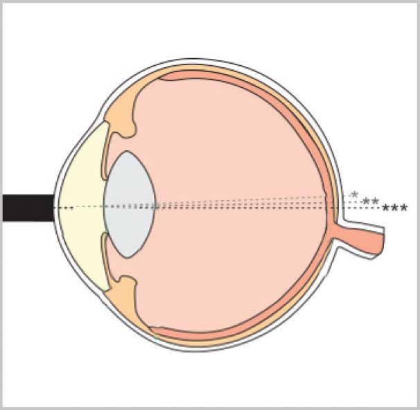
A unique grading algorithm automatically ranks the probe’s alignment along the axis of measurement. Alignment ranking is immediate, with each qualified measurement assigned a 1-star, 2-star or 3-star rank (3-star representing best alignment). Aided by the audible feedback, the user can adjust the probe’s contact angle during a procedure, correcting misalignments and thereby optimizing measurement.
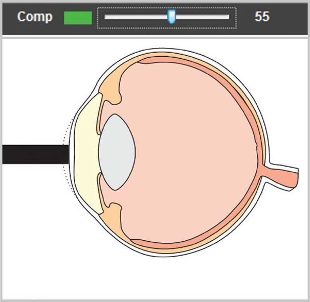
Unique compression lockout feature for use during contact measurements. When enabled, compression lockout will stop the system from measuring waveforms which show indications of corneal compression. Audible tones are provided to guide the user in adjusting contact pressure and aid in alleviating flattening of the cornea. The compression sensitivity level is adjustable to aid in obtaining contact measurements with minimum compression.
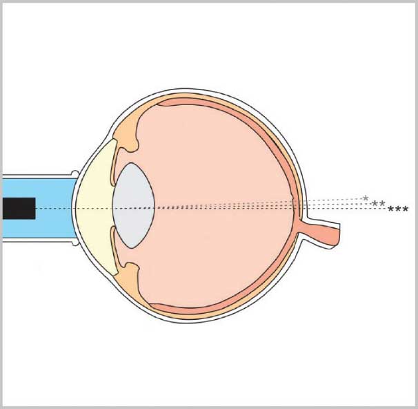
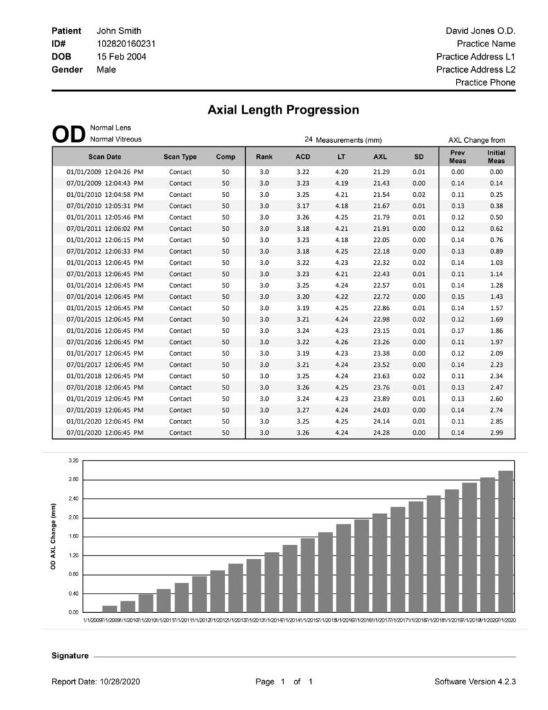
MONITORING AXIAL LENGTH PROGRESSION
The Flex A-Scan is tailored to meet the needs of your myopia management program. In addition to providing consistent, accurate and reliable axial length measurements, the Axial Length Progression Report charts the axial length changes of each patient over a period of time.
TOTAL IMAGING SOFTWARE SOLUTION
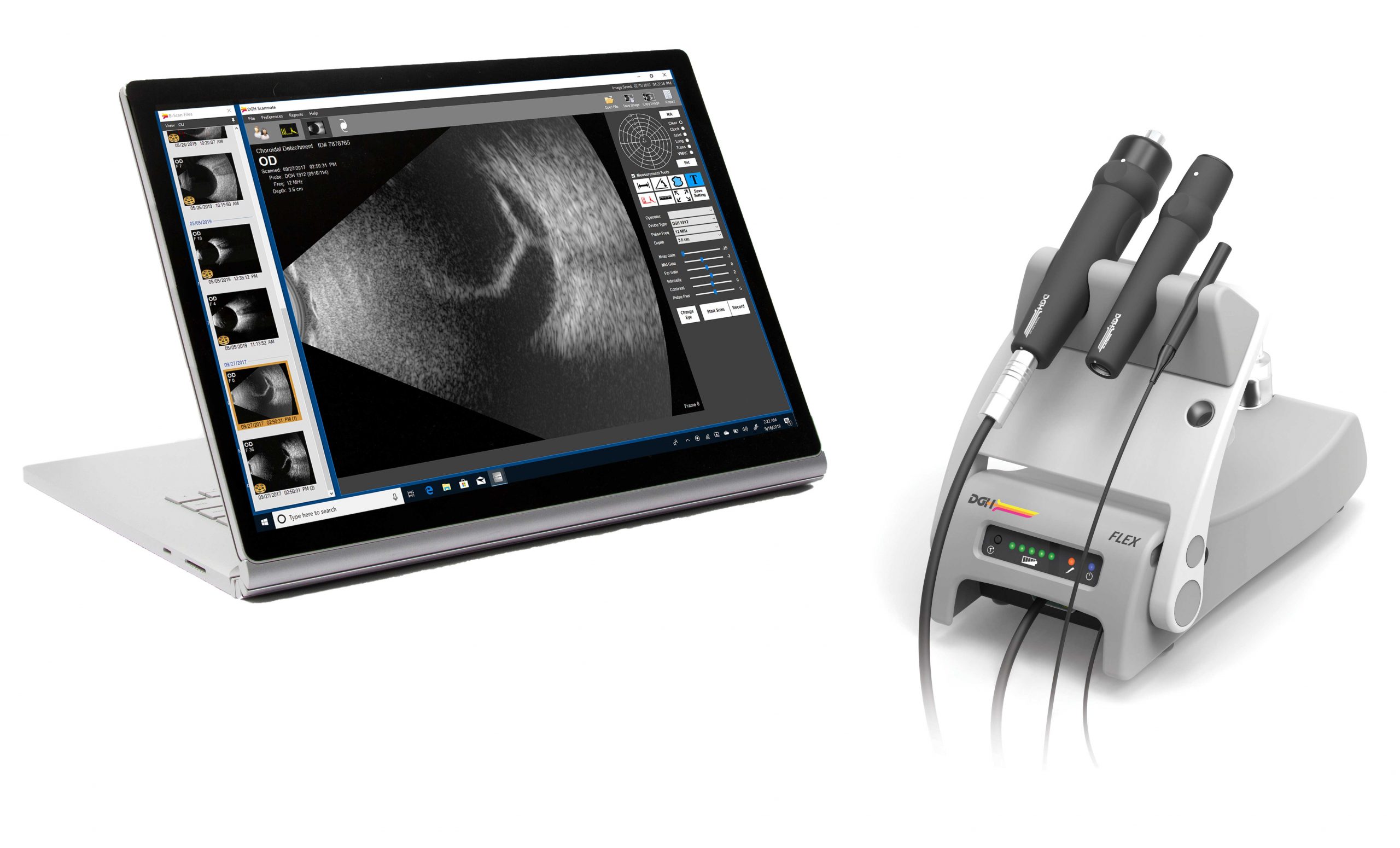
The Scanmate Flex combines the most advanced ultrasound technology available with the processing power, data storage and connectivity advantages of a personal computer. Patient data can be stored on a local computer, or in a centralized network where it can be accessed by multiple users. Patient records are fully searchable and can be exported in a format that is compatible with EMR/EHR systems. The Scanmate software is designed to work on a Windows® computer. The Scanmate Flex plugs into the USB 2.0 port of a Windows® computer that you may already have in your office or clinic*.
* See Specifications tab for minimum computer requirements.
The software was designed to accomplish an examination in 4 simple steps.
Step 1: Patient Data : Populate the necessary fields and you are ready to acquire images.
Step 2: Acquire Images: Click on desired modality icon, and start acquiring images and data.
Step 3: Image Processing and Quantification : The software offers tools to measure distance, area, and angles, and an annotation tool for notating images. Images can be enhanced by adjusting intensity, contrast, and gains (near,mid, and far).
Step 4: Reporting : The Scanmate Software offers a variety of report templates that summarize critical information and are print and .PDF ready.

|
B-Scan Specifications |
||
| B-Scan Probe Type | Internal, pivoting single-element transducer. 256 lines per scan. 60° scan angle. | |
| B-Scan Mode of Imaging | Contact | |
| Transducer Frequency Options | 12.5 MHz | 20 MHz |
| Lateral Accuracy | ± 550 μm | |
| Axial Accuracy (Theoretical) | 28.9 μm | |
| Focal Point (Nominal) | 20.00 mm | 21.00 mm |
| Depth of Field | 14.00 mm – 37.00 mm | 15.00 mm – 35.00 mm |
| UBM Specifications | ||
| UBM Probe Type | External (removable), pivoting single-element transducer. 256 lines per scan. 30° scan angle. | |
| UBM Mode of Imaging | Water Path (using ClearScan® probe cover) | |
| Transducer Frequency Options | 35 MHz | 50 MHz |
| Lateral Accuracy | ± 250 μm | |
| Lateral Resolution (Nominal) | 80 μm | 50 μm |
| Axial Accuracy (Theoretical) | 9.6 μm | |
| Axial Resolution (Nominal) | 65 μm | 50 μm |
| Focal Point (Nominal) | 13.00 mm | |
| Depth of Field (Nominal) | 11.50 mm – 14.00 mm | |
|
A-Scan Specifications |
||
| A-Scan Probe Type | Internal, fixed single-element transducer. | |
| A-Scan Mode of Measurement | Contact or Immersion (using Prager Shell® immersion shell) | |
| Transducer Frequency | 10 MHz | |
| Axial Length Measurement Range | 15.00 mm – 40.00 mm | |
| ACD Measurement Range | 2.00 mm – 6.00 mm | |
| Lens Thickness Measurement Range | 2.00 mm – 7.50 mm | |
| Resolution | 0.01 mm | |
| Repeatability | ± 0.03 mm STDEV (Immersion) | |
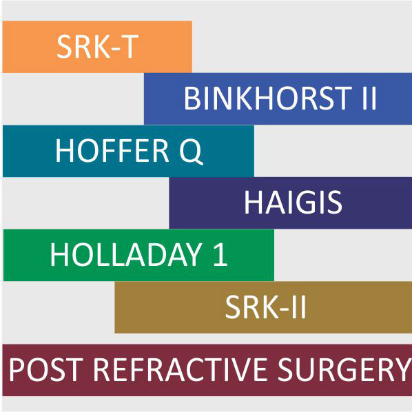
| IOL Formulas | SRK II, Binkhorst, SRK/T, Holladay 1, Hoffer Q, Haigis | |
| IOL Formulas (Post Refractive) | Double K (SRK/T), History Derived, Clinically Derived (Shammas), Refraction Derived, Contact Lens Over-Refraction | |
|
Base Unit Specifications |
||
| Dimensions | 209.55 mm (L) x 133.50 mm (W) x 101.60 mm (H) | |
| Weight | 8.00 lbs | |
| Battery Type | 3250 mAh Li-ion rechargeable battery (internal) | |
|
Software Application Requirements |
||
| Hardware Requirements | Intel i5 or higher, 4GB RAM or higher, 128 GB SSD/HDD or higher
2 x 2.0 USB, 1024 x 768 display resolution or higher |
|
| Operating System Requirements | Windows 8 or higher (32 or 64 bit), MS server 2008 R2 (64 bit),
MS Server 2012 / 2012 R2 (64 bit), MS Server 2016 (64 bit) |
|
DGH Technology, Inc.
110 Summit Drive, Suite B
Exton, PA 19341
Phone: (610) 594-9100
Fax: (610) 594-0390
Toll Free: (800) 722-3883
info@dghtechnology.com
SALES:
Eastern U.S.
Contact: Russell Schlage
(727) 692-8327
russell@dghtechnology.com
Western U.S.
Contact: Rafael Ramirez
(619) 993-4608
rafael@dghtechnology.com
International
Contact: Niket Dave
(312) 918 9808
nick@dghtechnology.com
Headquarters
Contact: Darrell Glasgow
(800) 722-3883
sales@dghtechnology.com
DGH DIRECT PURCHASE OPTION
Purchasing DGH products directly from DGH is the only way to ensure that you are purchasing the appropriate products for your practice at the best price possible. Simply contact DGH to arrange a personal consultation with our experienced Sales and Service Representatives who have the technical expertise required to determine which DGH products are best suited to your practice, and they will also discuss how the benefits of the Direct Purchase Option maximize the total value of your purchase.
BENEFITS OF THE DGH DIRECT PURCHASE OPTION
- Our Sales and Service Representatives are your personal DGH liaisons for all your pre-sales and post-sales needs.
- Special discount pricing for qualified buyers on all DGH products.
- On-line demonstrations.
- Customer Satisfaction Guarantee: Return any purchase within the first 30 days for a full refund.
- Discounted pricing for a new DGH product when trading in a used DGH product.
- Special extended warranty offers.
- Priority Customer Service.
- Customized financial options available through DGH affiliates.
Note: The DGH Direct Purchase Option is available only for customers based in the USA.


